Dental X-Rays Service
X-rays are also known as radiographs, they are an essential part of any dental treatment program. They are diagnostic, but they can also be preventative, by helping a dentist to diagnose your potential oral care issues in your mouth before they become a major problem. An x-ray is a type of ray energy that passes through soft tissues and is absorbed by dense tissue. Your teeth and bones are very dense, so they absorb X-rays, while X-rays pass more easily through your gums and cheeks.
X-rays are divided into two main categories, Intraoral and Extraoral where intraoral is an X-ray that is taken inside the mouth while an extraoral X-ray is taken outside of the mouth.
Intraoral X-rays are the most common type of X-ray taken in dentistry. They give a high level of detail of the tooth, bone and its supporting tissues of the
mouth. These X-rays:
• Find cavities
• Look at the tooth roots
• Checks the health of the bones around the tooth
• Determine if there is any periodontal disease which is becoming an oral care issue
• See the status of developing teeth
• Otherwise, monitor good tooth health through prevention
Types of Dental X-rays
The two types of Dental X-rays: intraoral (the x-ray film is inside the mouth) and extraoral (the x-ray film is outside the mouth).
Intraoral x-rays are the most common type of x-ray which shows the structure of the inside mouth. There are many types of intraoral x-rays:
• Bite-wing x-rays shows the details of the upper and lower teeth in one area of the mouth. Each bite-wing shows a tooth from its
crown to the level of the supporting bone. Bite-wing x-rays detect the tooth decay and also changes the thickness of bone caused by gum diseases. Bitewing x-rays
can also help to determine the proper fitting of the crown (a cap that completely covers a tooth) or other restorations (e.g., bridges). It can also see any
breakdown of the dental fillings.
• Periapical x-rays show the whole tooth — from the crown to the root where the tooth attaches into your jaw. Each periapical x-ray
shows all teeth in one portion of either the upper or the lower jaw. Periapical x-rays detect any unusual changes which should not happen, in the root and even
surrounding bone structures.
• Occlusal x-rays track the development and placement of an entire arch of your teeth in either the upper or the lower jaw.
Extraoral x-rays are used to detect the dental problems in the jaw and the skull. There are many types of extraoral x-rays:
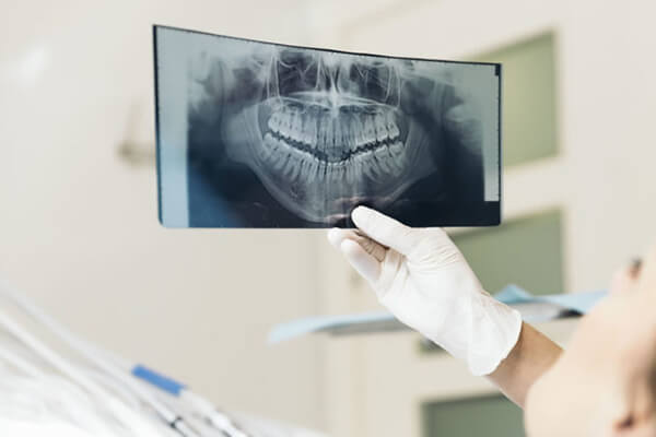
• Panoramic x-rays shows the entire mouth area — all the teeth of both the upper and lower jaws are shown — on a single x-ray.
This x-ray detects the position of fully emerged as well as emerging teeth, can see impacted teeth and helps in diagnosing tumors.
• Tomograms show a particular layer of the mouth and blur out the other layers. This x-ray examines the structures that are difficult to be seen clearly
because other nearby structures are blocking that view.
• Cephalometric projections show an entire side of the head. This x-ray looks at the teeth and the jaw relation.
• Another test that uses x-rays is called a sialogram. This test uses a dye, which is injected into your salivary glands so they can be seen on x-ray film.
We might order this test to look for salivary gland problems.
• Dental computed tomography (CT) is a type of imaging that shows an interior structure in 3-D. This type of imaging is used to find the problems in the bones
of the face such as tumors, cysts, and fractures.
• Cone Beam CT is a type of x-ray that creates 3-D images of your dental structures, soft tissues, nerves, and the bones. It helps us to guide tooth implant
placement and evaluates cysts and tumors in the mouth and the face. It can also detect problems in your gums, roots of the teeth, and the jaws. Cone beam CT is
just like the regular dental CT, they both give accurate and high-quality images. The cone-beam CT machine rotates around the patient’s head, it captures all
data in one rotation. This method also exposes patients to a higher level of radiation. A unique advantage of cone beam CT is that it can be only used in a
dentist’s office. Dental computed CT equipment is only available in hospitals or on the imaging centers.
• Digital imaging is a 2-D type of dental imaging that allows the images to be sent directly to a computer. The images can be viewed on screen or can be
stored and can be printed out in a matter of seconds. Digital imaging has several other advantages in comparison to other traditional x-rays. The image taken
of a tooth, for example, can be enhanced or enlarged. This makes it easier for us to see the tiniest changes that can’t be seen in an oral exam.
• MRI imaging is an imaging method that takes a 3-D view of your oral cavity including the jaw and teeth.
Why Use a Panoramic X-ray?
Just because a panoramic X-ray shows the entire mouth in one picture, it doesn't produce the exact details needed to show the cavities. This type of X-ray does, however, show problems such as bone abnormalities and fractures, impacted teeth, cysts, infections, and tumors. If any of these problems has been suspected, then we will choose to take a panoramic X-ray. We may also use this imagery method when planning for treatments such as braces, implants, and dentures.
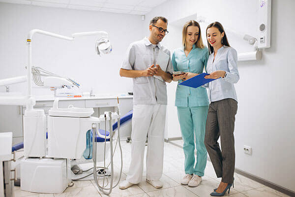
Welcome To ALL Brite Dentistry
We at All Brite Dentistry want to give each of our patients the most gentle and highest quality dental care possible. It starts with your very first phone call. Compassion and understanding are a priority.
Read MoreAppointment
Book An Appointment Now!
We at All Brite Dentistry want to give each of our patients the most gentle and highest quality dental care possible. It starts with your very first phone call. Compassion and understanding are a priority.
Our Services
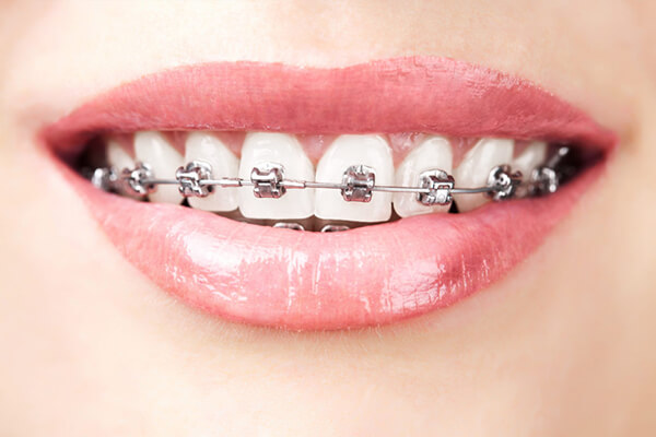
Teeth braces or dental braces
Dental braces are also known as braces, or orthodontic cases are the devices which are used in orthodontics that aligns and straightens your teeth.
Read More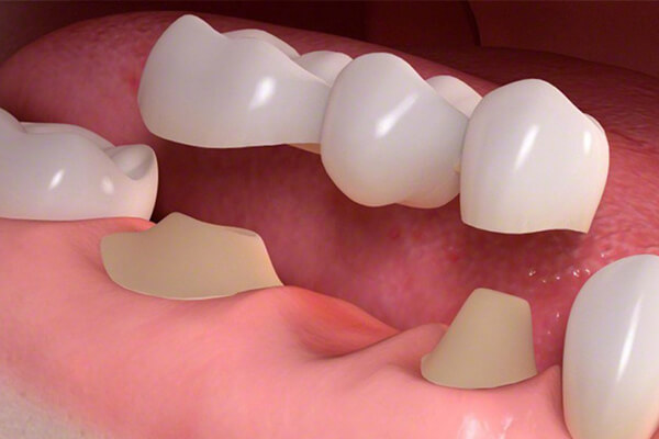
Dental bridges
A bridge is a fixed dental restoration which usually replaces one or more missing teeth by joining an artificial tooth adjacent to the other teeth.
Read More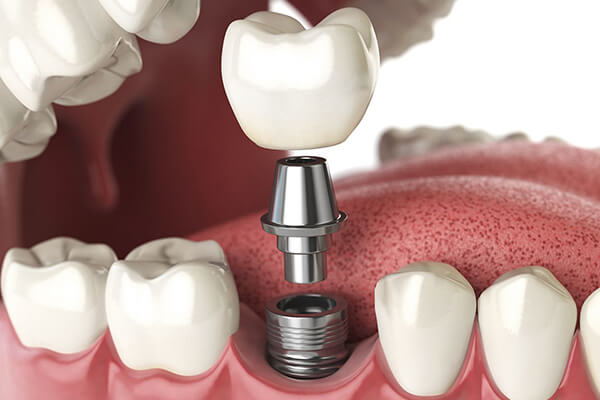
Dental implants and restorations
A dental implants are also known as an Endosseous implant or a fixture is a surgical component that interfaces with the bone of the jaw or skull.
Read More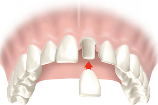
Dental crowns or teeth crowns
A crown, which is also known as a dental cap, is a type of dental restoration which completely caps, covers or encircles a tooth or dental implant.
Read More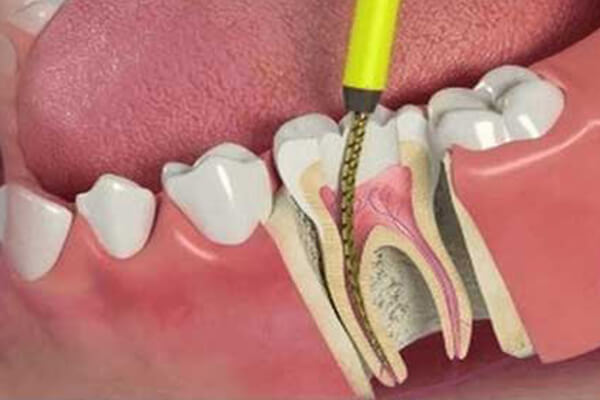
Root canal treatment/procedure
Endodontic therapy, which is also known as endodontic treatment or root canal therapy, is a treatment for the infected pulp of a tooth which results in the cure of infection.
Read More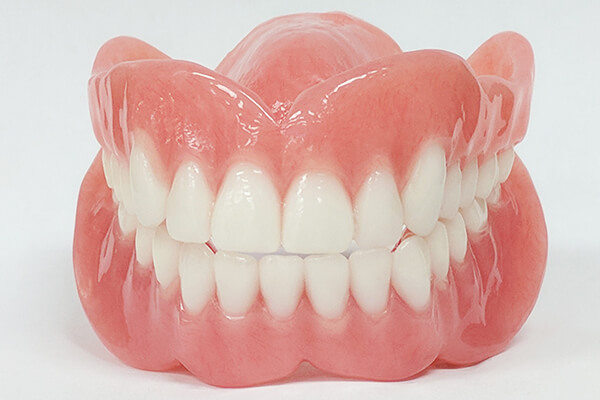
Dentures
Dentures are the artificial teeth worn by the people who don't have their real teeth. Dentures are also called false teeth or dental plate.
Read More
Teeth extractions
Although permanent teeth were meant to last for the lifetime, there are a number of reasons because of which tooth extraction may be needed.
Read More
Dental hygiene
Dental Hygienics is one of the leading providers of dental decontamination equipment and products.A dental hygienist is a licensed dental professional.
Read More
Teeth veneers
Dental veneers are wafer-thin, customized shells of tooth-colored materials designed to cover the front surface of your teeth to improve your appearance.
Read More
Teeth whitening
Teeth whitening involves the bleaching of your teeth to make them lighter, clean and white. It can't make your teeth brilliantly white, but it can lighten the existing color.
Read More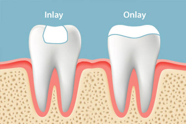
Inlays and onlays
Inlays and Onlays are used when the tooth has experienced too much of damage that it cannot support a basic filling, but not so much damage that a crown is necessary.
Read More
Cosmetic dental fillings
Aesthetic dentistry is a branch of dentistry that involves skills and techniques to improve your alignment, smile’s appearance, shape, color, and size.
Read More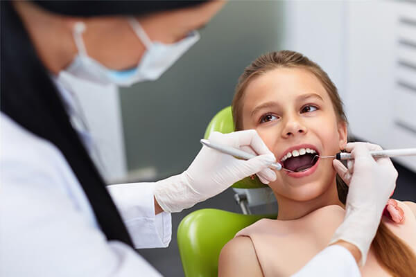
Pediatric dentist
Pediatric dentists are also called kids’ dentists or child dentists. They are basically dedicated to the oral health of children from infancy through their teenage.
Read More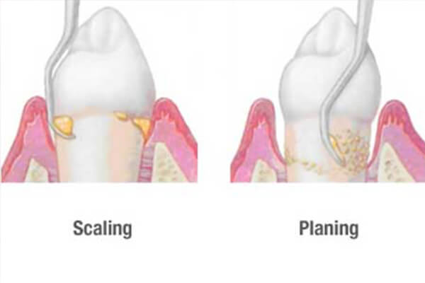
Teeth scaling and root planning
Teeth scaling and root planning is a procedure involving removal of dental plaque and calculus ( tartar ) and then smoothing of the (exposed) surfaces of the roots.
Read More
Periodontal gum disease
Periodontology is the specialty of dentistry that studies for the supporting structures of teeth, as well as the diseases and conditions that can affect them.
Read More
Cosmetic dentistry
Cosmetic dentistry refers to any kind of dental work that improves the appearance (not necessarily the functionality) of teeth, gums and/or bite.
Read More
Family dentistry
Consetetur sadipscing elitr, sed diam nonumy eirmod tempor invidunt ut labore et dolor erat, sed diam voluptua, maiores possimus fugiat repellat totam.
Read More
Emergency dentistry
Before searching for an emergency Dentist, It is important to know that what kind of injuries really required an emergency dental care, so you can make sure that teeth are taken care.
Read More
Dental exams and cleanings
Many people feel afraid of teeth cleanings. Between the prodding, strange noises, and the occasional jaw discomfort, it’s easy to understand their apprehension.
Read More
Dental X-Rays
X-rays are also known as radiographs. An x-ray is a type of ray energy that passes through soft tissues and is absorbed by dense tissue. They are diagnostic,also be preventative.
Read More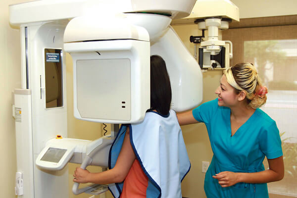
CT scan
CBCT stands for Cone Beam Computed Tomography. It is a technology used to take three dimensional (3-D) images of your teeth, maxillary sinus, nerve pathways, and bone .
Read More
Night guards
A mouth guard is a protective device for your mouth that covers your teeth and your gums to prevent and reduce the injury to your teeth, arches, lips, and your gums.
Read More
Sleep apnea dentist
Sleep apnea is a sleep disorder in which your breathing starts and stops repeatedly. If you snore loudly and feel tired even after having a full night's sleep.
Read More
Dental botox theraphy
The active, therapeutic neurotoxin is harvested, separated and purified in a laboratory to make it safe and effective for the treatment.
Read More